Structure & Function of Bone Copy

Introduction to this section

In this section you will learn more about how bone behaves as a physiological tissue in response to forces. You should gain a basic appreciation of how bone tissue is arranged to be both strong and dynamic and protects against excessive forces. Knowing how a ‘normal’ bone responds to stresses helps us appreciate what happens when bone tissue becomes ‘abnormal’ and no longer responds the same way to even the smallest forces and fractures result.

In Your Manual

Basics: Page 159-165 | Clinical: Page 175-180

Bone as Material

Over a few centuries, bone structure and function has been discussed and studied however, significant findings did not occur until the late 1600s starting with Leeuwenhoeck (1674) who was the first to publish observations of osteons (building blocks of bone) at the microscopic level. Landmark findings included the, the concept that bone is formed and resorbed throughout life (Munro, 1776), the finding that the surrounding muscles determine the form of bone (Fick, 1856) and the statement that there is a relationship between function and structure of bone (Von Meyer, 1867).


It is this theme of the related structure and function that guided the research in the next century with important discoveries and theories such as proposed laws by Wolff (1870) that summarize an interdependence between form and function of bone, and the proposal by Roux (1883) that the orientation of bone trabecula corresponds to the direction of tension/compression which is an example of how bone architecture follows engineering principles.


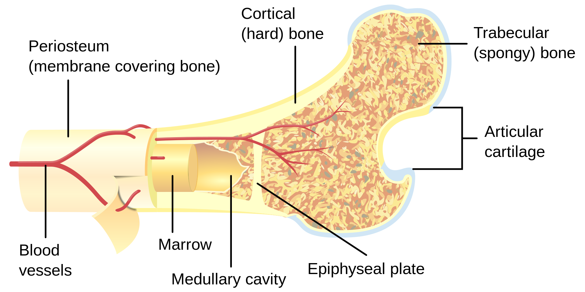
Bone is a living tissue composed of a protein matrix which is osteocollagenous and minerals (calcium salts). Twenty percent of the bone is water, while the matrix and salts contribute 35% and 45% respectively. Bone can be classified as cancellous (otherwise known as trabecular) or cortical (compact) which do not differ greatly biomechanically despite their differing density and porosity.

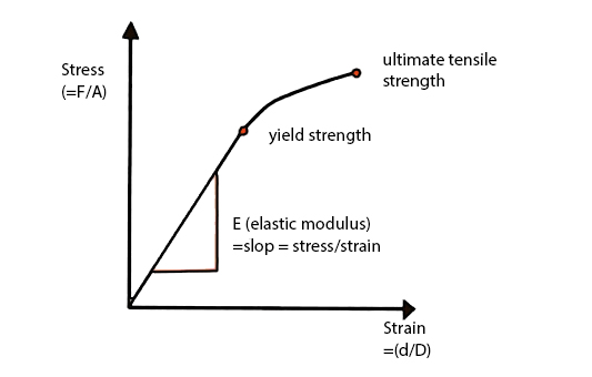
stresstraincurvev2

Stress: force per unit area

Strain: change in length relative to original length

Bone as a Material  

Bone can be examined as a material - and the material properties of the bone can be characterized by the load- deformation relation. By applying a load to a tissue such as bone, then the change in length (deformation) can be measured.

Intrinsic material properties of bone can be expressed in two other measures:

Stress: force per unit area

Strain: change in length relative to original length

Stiffness of the bone is the relationship between stress and strain and this relationship can be plotted like a graph (see figure 2.1).  Each region of the slope provides a reflection on the bone’s material properties:

Elastic/linear region: an area of low stiffness, (calculation of this relationship provides a number =elastic modulus). This is the region in which changes in the original length of the material will return to normal length. On figure 2.1 this is the region which is linear.

Plastic region: high stiffness region – where the material yields and damage accumulate here due to the rearrangement of internal structures. On figure 2.1, this is the non-linear (curved) region of the graph.

Ultimate yield point - Following the plastic region where the material 'breaks'.

The area under the curve indicates the amount of energy absorbed upon application of force.

Bone as a structure

Bone can also be characterized by its structure. Unlike the material properties of bone, structural properties are dependent on the geometry of the bone. Size, shape, cross-sectional area, and trabecular orientation in the body (ie., direction that it encounters compressive loads) determine the biomechanical properties. The stress that a bone can withstand depends significantly on the size (cross-sectional area) of that bone and therefore those bones whose function is to withstand large stresses must have a large cross-sectional area -right back to the connection between structure and function!

FACT: most asymptomatic bones can withstand forces between two and five times what they normally are subjected to in activities of daily living.
Mechanical Behavior of Bone

https://www.google.com/url?sa=i&url=https%3A%2F%2Fimgur.com%2Fbeta%2Fdisable%3Fredirect%3D%252Fgallery%252FSC12w4d%252Fcomment%252F628488472%253Fnc%253D1&psig=AOvVaw1OSlno79hQht-V2PNuy091&ust=1604172901439000&source=images&cd=vfe&ved=0CA0QjhxqFwoTCLCHqueH3ewCFQAAAAAdAAAAABAD

Source: Imgur

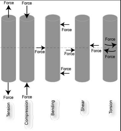
You can certainly imagine that your daily activities result in all kinds of forces placed on the body - every change of position results in some type of force being transmitted through your bones and joints. Therefore, bones are subjected to compressive, tensile and shear stresses daily. Occasionally external forces are applied to our bones which are excessive, take for example being hit on the leg by a taxi while trying to cross the street. This could result in a bending load being applied to your tibia. See figure below for what happens on each side of the bone.

Fracture Patterns

 Fracture Pattern: Bending Force

So we know that if a bending force is applied to a long bone, the failure point is more than likely to be the convex (tension) side. This results in tension failure to occur progressively across the bone creating a transverse fracture. There is a purpose for the bone to fracture - it is the method in which the energy applied during the trauma is dissipated. Therefore it is logical that if a high velocity force is applied to the bone, more energy needs to be dissipated and more cracks in the bone would be generated in the bone to dissipate this energy. Low velocity impacts usually result in a single transverse crack extending 1/3-1/2 of the circumference of the bone, often making unpredictable, oblique angles.

Fracture Pattern: Torsion

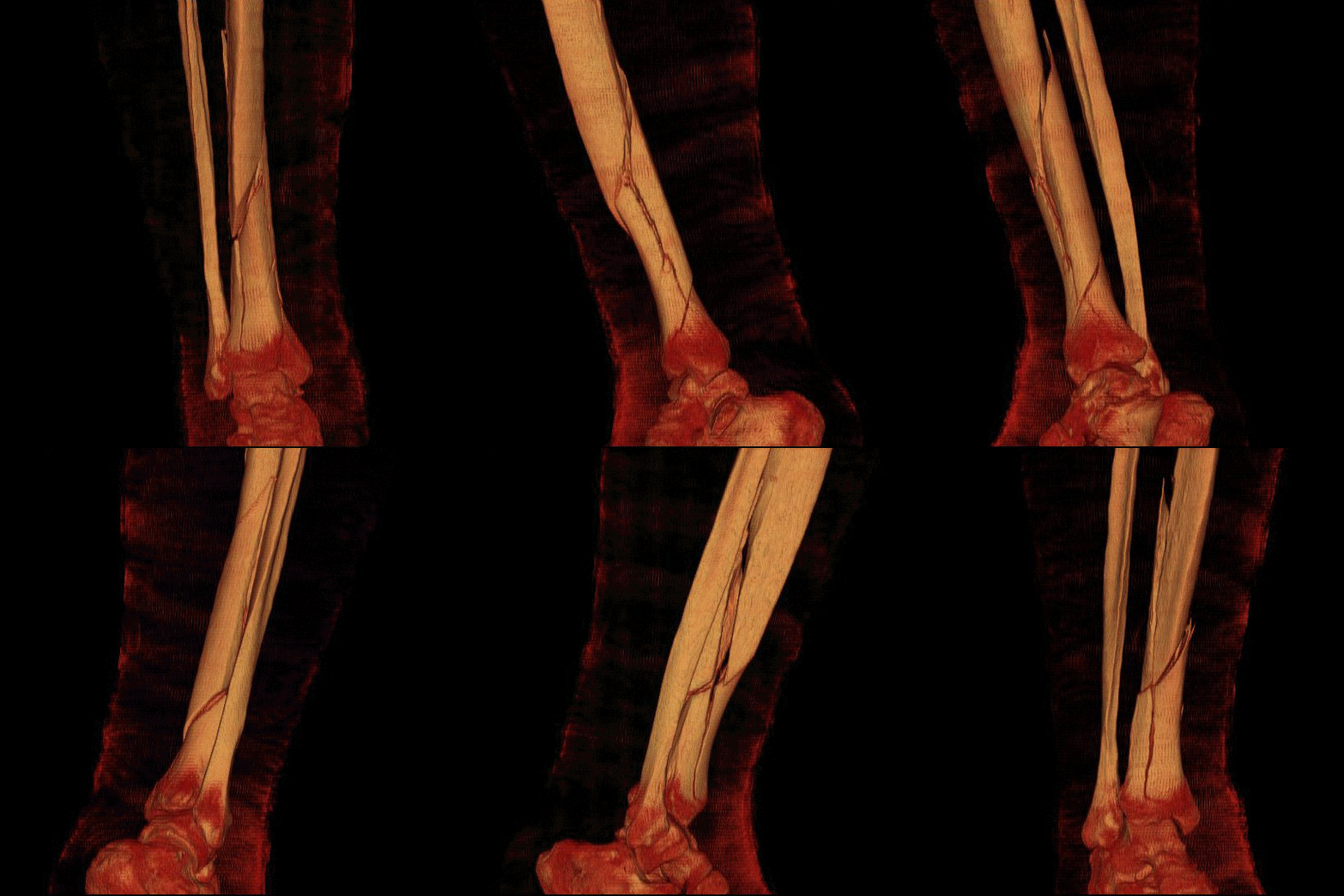
Of course injuries to the bones don't always occur by bending but by twisting forces, such as can happen when a foot is fixed and the body falls or rotates forcefully, for example a fall while skiing. The resulting torsional load on the tibia in this case produces a constant moment throughout the long bone since the moment applied to the fixed segment (in the ski boot) is the exact magnitude but in the opposite direction as the moment closer to the knee joint.

Generally the pattern of the torsional fracture is thus a spiral which is perpendicular to the maximum tensile strength applied. Similar to the bending fracture pattern, loads that are rapidly applied produce more fracture lines - a double spiral pattern.

Fracture Pattern: Compression

Compressive loading involves the application of equal and opposite loads into the structure, resulting in compressive stress and strain within the structure and a resulting shortening and widening of that structure.  If the load is applied on the plane perpendicular to the object surface, this results in maximal compressive stress. As the vertebrae are subjected to high compressive loads under normal circumstances, it is expected that these bones in particular would be susceptible to undergoing compressive fractures. In other joints, compression fractures can be elicited with abnormally high muscular contraction forces around the joint.  Although this doesn’t typically apply to a vertebral segment, consider the effect of repeated, high muscular work on one aspect of the vertebrae and that contribution to developing vertebral fractures23.

Figure 1 - Vertebral Wedge Fracture

Typical patterns of vertebral fractures include crush and wedge fractures. Crush fractures occur in the condition in which compressive forces are distributed nearly equally throughout the entire vertebral body,  thus causing an equal loss of height throughout the entire vertebral body.  Wedge fractures, on the other hand, are generally a result of accentuated compressive forces on one aspect (most typically anterior) of the vertebral body, resulting in a more wedge shaped vertebra. (see Figure 1)

Effects of Aging on Bone

Cortical Bone:

The affect of aging results in cortical bone’s diminishing ability to resist fracture. Elastic properties decrease somewhat but it is the toughness and strength of the bony material which diminishes even more so.  Research has revealed that the stiffness of femoral cortical bone reduces 1-2% per decade after 35 years old, while maximum stress (strength) decreases 2-5% per decade (Burstein, 1976; Zioupos, 1998). Most significantly the toughness of cortical bone (the energy to fracture) decreases 10% per decade which means that lower energy events impacting the bone in the older persons would result in a fracture which would not occur similarly in a younger person.  In addition, cortical bone is porous to begin with and this porosity increases significantly with age (McCalden, 1993).

Trabecular Bone

Trabecular, or cancellous, bone strength is dependent largely upon the density of its trabeculae – according to some research, this accounts for approximately 60% of the variance in strength in sites around the body (Goulet, 1994). Since trabeculae are arranged in different patterns within bone, if forces are directed through the bone trabeculae along its’ principal orientation, strength variance rises to 90%.

With aging in both men and women, the apparent density of trabecular bone decreases significantly and thus, the strength of the bone is proportionately decreased (McCalden, 1997; Mosekilde, 1987). Its been reported that the compressive strength of vertebral trabecular bone decreases between the ages of 25-75 year by 70% and proximal femoral trabecular bone loses 50% of its strength in the same time span (McCalden, 1997). Accounting for this loss of strength is a reduction in the thickness and number of individual trabeculae, resulting in a loss of density (Goldstein, 1993; Goulet, 1994).


Bone_cross-section

Source: Wiki2

Whole Bone Changes with Age:

With age, the proximal femur undergoes significant loss of structural integrity, thus leaving it vulnerable to damage under low force conditions.  Research comparing younger and older femoral specimens revealed that in the older specimens there was a reduction of strength by 50%, stiffness by 30% and overall toughness by 70% (Courtney, 1995).


Similarly, vertebrae also undergo age-related changes in strength which are particularly related to the density and strength of the vertebral trabecular bone.  Between the ages of 30 and 90, compressive strength of the vertebrae decreases by 80% (Ebbesen, 1999).

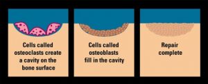
Bone is living tissue, constantly renewing itself. Although bone is strong and relatively flexible, everyday wear and tear causes tiny structural defects, much like those that occur in the foundations of a building over time. In our bodies, osteoclasts and osteoblasts are two groups of specialized cells that perform the work of a "maintenance crew.“ 

As we age, the two groups of cells that form the maintenance crew become less efficient in working together - the osteoclasts remove old bone faster than the osteoblasts are able to rebuild it. 

Studies of older adults show that adequate calcium intake can slow bone loss and lower the risk of fracture.

Burstein, A. H., Reilly, D. T., & Martens, M. (1976). Aging of bone tissue: mechanical properties. J BoneJoint Surg Am, 58(1), 82-86.

Courtney, A. C., Wachtel, E. F., Myers, E. R., & Hayes, W. C. (1995). Age-related reductions in the strength of the femur tested in a fall-loading configuration. J Bone Joint Surg Am, 77(3), 387-395.

Ebbesen, E. N., Thomsen, J. S., Beck-Nielsen, H., Nepper-Rasmussen, H. J., & Mosekilde, L. (1999). Age- and gender-related differences in vertebral bone mass, density, and strength. J Bone Miner Res, 14(8), 1394-1403.

Fick, A. (1856). Medizinische Physik. Supplementband in Müller-Pouillet's Lehrbuch der Physik fürMediziner

Goldstein, S. A., Goulet, R., & McCubbrey, D. (1993). Measurement and significance of three- dimensional architecture to the mechanical integrity of trabecular bone. Calcif Tissue Int, 53 Suppl 1, S127-132; discussion S132-123.

Goulet, R. W., Goldstein, S. A., Ciarelli, M. J., Kuhn, J. L., Brown, M. B., & Feldkamp, L. A. (1994). The relationship between the structural and orthogonal compressive properties of trabecular bone. J Biomech, 27(4), 375-389.

Leeuwenhoek, A. (1674). Microscopical observations concerning blood, milk, bone, the brain, spittle, and cuticula, etc. Phil Trans, 9, 121-128.

McCalden, R. W., McGeough, J. A., Barker, M. B., & Court-Brown, C. M. (1993). Age-related changes in the tensile properties of cortical bone. The relative importance of changes in porosity, mineralization, and microstructure. J Bone Joint Surg Am, 75(8), 1193-1205.

McCalden, R. W., McGeough, J. A., & Court-Brown, C. M. (1997). Age-related changes in the compressive strength of cancellous bone. The relative importance of changes in density and trabecular architecture. J Bone Joint Surg Am, 79(3), 421-427.

Monro, A. (1776). Anatomy of the humane bones. (10 ed.). Dublin.

Mosekilde, L., & Danielsen, C. C. (1987). Biomechanical competence of vertebral trabecular bone in relation to ash density and age in normal individuals. Bone, 8(2), 79-85.

Notelovitz, M., Ware, M., & Tonnessen, D. (1998). Stand tall!: Every woman's guide to preventing and treating osteoporosis. Gainesville, FL: Triad Pub.

Roux, W. (1883). Beiträge zur Morphologie der funktionellen Anpassung. Archiv für Anatomie und Entwickelungsgeschichte., 76–162.

Von Meyer, H. (1867). Die Architecktur der Spongiosa. Reichert und Dubois-Reymond’s Archiv, 34:615-28.

Wolff, J. (1870). Ueber die innere Architektur der Knochen und ihre Bedeutung für die Frage vom Knochenwachstum. . Arch. Pathol. Anat. Physiol. Klin. Med. Virchows Arch., 50, 389-453.

Zioupos, P., & Currey, J. D. (1998). Changes in the stiffness, strength, and toughness of human cortical bone with age. Bone, 22(1), 57-66.

Back
Subscribe